Projets
Equipe Destin Cellulaire

Mise en place du tube neural larvaire chez l'ascidie

 As well as the neural cells which come from the vegetal A-line cells from a cell fate choice between neural and notochord (see project 1), CNS precursors are also specified in the animal cells (a-line and b-line) following cell fate decisions between neural and epidermal fates.
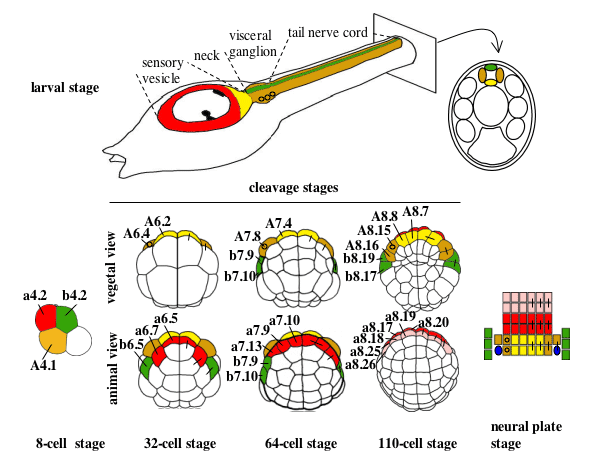
Figure 1. Cell lineages of the ascidian larval CNS (from Hudson and Yasuo, 2005). Cell lineages are indicated as follows: the a-line is coloured red (anterior sensory vesicle precursors) or pink (anterior epidermis and pharynx/neurohypothesis precursors); b-line is green and A-line yellow at the 8-cell stage and light yellow (medial cells) or tan (lateral cells) from the 32-cell stage. Bars connecting two blastomeres on the right-hand side of the drawings indicate sister cell relationship.Small circles in the lateral-A-line precursors on the left hand side indicate that these blastomeres give rise to the motoneurones; the final position of the motoneurones in the visceral ganglion is also indicated on the drawing of the larvae. At the neural plate stage, only the neural plate is shown and the dark blue ovals represent the secondary muscle lineage.

In order to form a fully developed CNS, the neural cells, once generated, obtain positional information which govens patterning along the dorsal-ventral and anterior-posterior axis. This leads to the generation of specific cell-types in the CNS, such as the motoneurones which form in the ventral-lateral part of the visceral ganglion (see Fig.1). Patterning of the neural cells begins as early as the early gastrula stage, when we first see a difference in the genes expressed between the medial and the lateral A-line neural precursors (Fig.2).
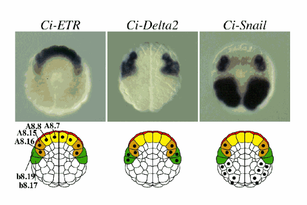
Figure 2. Differential expression of Ci-Snail and Ci-Delta2 in lateral but not medial A-line neural precursors. On the right is a picture of an in situ hybridization with a probe for Ci-ETR,a general neural marker, which is expressed in all 8 A-line neural precursors at the early gastrula stage. Expression of Ci-ETR in the lateral-most A-line neural precursor (A8.16) is weaker than the other A-line neural blastomeres, consistent with the fact that A8.16 is not yet fate restricted to neural fate, but still contains muscle fate. In contrast to Ci-ETR, Ci-Snail and Ci-Delta2 are specifically expressed in the lateral neural precursors (A8.15 and A8.16) and not the medial precursors (A8.7 and A8.8). Expression of these two genes is the earliest known molecular difference between medial and lateral neural precursors. This suggests that, at least by the early gastrula stage, medial and lateral A-line neural precursors start to undergo a different molecular program. Below each picture is a schematic representation of an embryo at the early gastrula stage. Different lineages are shown with the same colour code as in the Cell lineages of the ascidian larval CNS figure. A spot marks the cells expressing the gene analysed in the panel above. For more details see Hudson and Yasuo, 2005.

By neural plate stages, expression of genes can be found in specific anterior-posterior as well as dorsal-ventral domains.
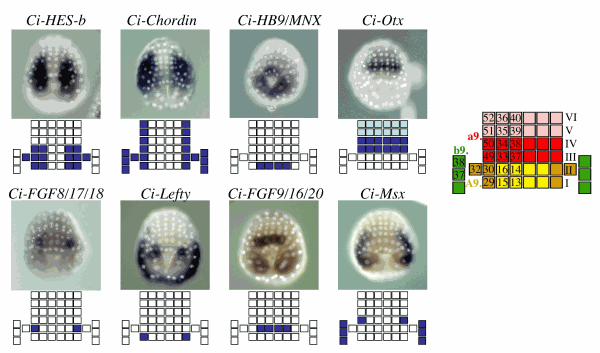
Figure 3. A collection of neural plate markers. At the right of the panels is a schematic drawing of the neural plate when it consists of 6 rows of cells (mid-gastrula stage), with each square representing a neural plate cell. Names of each cell are indicated, which should be prefixed with an ‘a9.’ for a-line cells, ‘A9.’ for A-line cells and ‘b9.’ for b-line cells. I-VI indicate the row number with Row I the closest to the blastopore, which is at the most caudal position of the neural plate. The colour scheme used is the same as in the Cell lineages of the ascidian larval CNS figure. The gene expression pattern analysed is indicated above the photo of the embryo. In these in situ hybridizations the nuclei have been labelled to allow easier identification of individual cells. A schematic drawing of the neural plate with the blastomeres expressing each marker coloured in blue is presented below each embryo. Some of these expression patterns are published in Hudson and Yasuo, 2005; Ci-Otx expression pattern is published in Hudson and Lemaire, 2001.

We are analysing cell-cell interactions, cell signalling pathways and transcriptional gene regulation to try to understand how the patterning of the neural plate is generated



Personnels Enseignants

Photo annuaire
Carine Barreau
Chercheur | SORBONNE UNIVERSITE
carine.barreau[at]imev-mer.fr
+33 (0) 4 93 76 39 73
Bâtiment Jean-Maetz
Photo annuaire
Joao Carvalho
Chercheur | SU
joao.carvalho[at]imev-mer.fr
+33 (0) 4 93 76 37 89
Station Zoologique

Master Biologie Moléculaire et Cellulaire

Parcours Biologie Cellulaire et du Développement & Cellules Souches

DEVELOPPEMENT DES ORGANISMES MARINS (DOMO)

Cette UE se déroule sur 2 semaines et a lieu au laboratoire de Biologie du Développement de Villefranche -sur-Mer (LBDV). Elle inclut l’examen de l’UE d’analyse scientifique (5V089) suivie par les étudiants de la spécialité de Biologie du Développement.

Durant la 1ère semaine, les étudiants participent à des ateliers et des rencontres avec les chercheurs du laboratoire.

Durant la 2ème semaine, les étudiants sont répartis dans les équipes pour y réaliser un mini projet qu’ils présentent le dernier jour du cours. Le cours est donné en anglais pour tout ou partie.

Voir la fiche UE

Modalités  d’évaluation

Présentation orale du mini-projet (binôme, 100 %)  

UE en anglais (partiellement ou totalement)

Master BMC, S3, 6 ECTS

Code UE: 5V200

Responsable de l’UE: Carine BARREAU (MCU): carine.barreau [at] obs-vlfr.fr

Master Biologie Intégrative & Physiologie

Parcours Biologie et Bioressources Marines (BBMA)

ORGANISMES MARINS & MODELES BIOLOGIQUES

Cette UE permet aux étudiants de 1ère année de Master de passer 2 semaines à l’Observatoire Océanologique de Villefranche-sur-Mer. Le cours est obligatoire pour les étudiants du Master Biologie Intégrative, parcours Biologie et Bioressources Marines (BBMA) tandis qu’il peut être choisi en option par les étudiants du Master Biologie Moléculaire et Cellulaire (BMC). Les étudiants participent à des ateliers de présentation des organismes marins utilisés par les équipes de recherche du laboratoire (LBDV) et apprennent à les manipuler au cours de travaux pratiques dont les thématiques vont de la Biologie du Développement fondamentale à la toxicologie appliquée.

Voir la fiche UE

Modalités  d’évaluation

Analyse d’article et présentation orale (50%)

Compte-rendu écrit de TP (50%)  

UE partiellement en anglais

Master BIP, S2, 6 ECTS

Code UE: 4B022 (ouverte au Master BMC)

Responsable de l’UE: Carine BARREAU (MCU): carine.barreau [at] obs-vlfr.fr

Licence de Sciences de la Vie

Parcours Biologie et Bioressources marines

Biologie Des Organismes Marins Et Diversité Des Recherches

Cette UE complémentaire se déroule sur 2 semaines et permet aux étudiants de découvrir les différents aspects (métiers & recherche) de l’Observatoire Océanologique de Villefranche-sur-Mer (OOV). Ateliers et journal clubs sont organisés afin que les étudiants mettent en pratique leurs connaissances théoriques en biologie et développent leur capacité de communication scientifique en français et en anglais.

Voir la fiche UE